Why Echogenic Embryo Transfer Catheters Are Becoming Essential for Ultrasound-Guided IVF Procedures
Introduction
Embryo transfer is widely recognized as one of the most critical steps in the in vitro fertilization (IVF) process. Even when high-quality embryos are available, the success of treatment depends on accurate and atraumatic embryo placement within the uterine cavity.
Over the past decade, ultrasound-guided embryo transfer has become the preferred approach in many fertility clinics because it enables physicians to visualize catheter placement in real time and improve procedural precision. However, the effectiveness of ultrasound guidance depends not only on the imaging system but also on the visibility of the catheter itself.
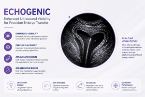
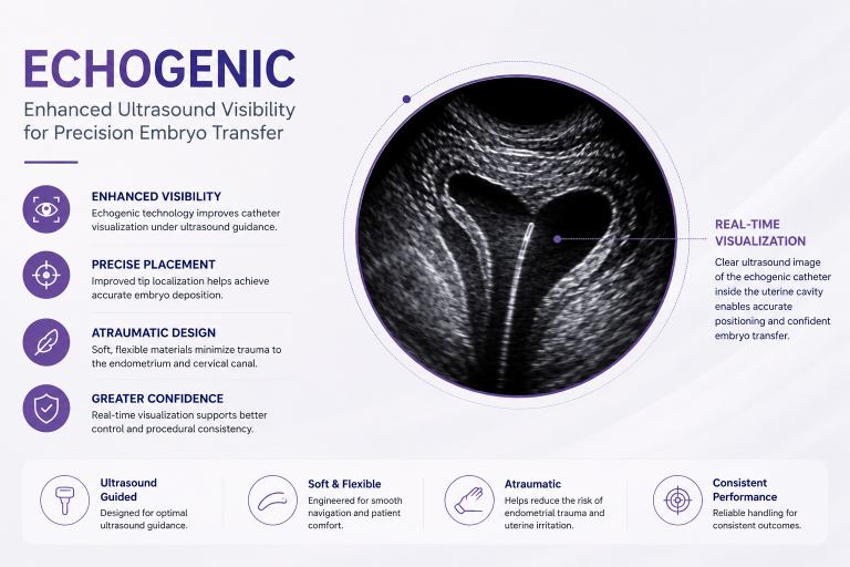
This is why the echogenic embryo transfer catheter has become an increasingly important innovation in reproductive medicine. By improving catheter visibility under ultrasound, echogenic technology supports more accurate embryo placement, reduces unnecessary manipulation, and enhances physician confidence throughout the transfer procedure.
Why Catheter Visibility Matters During Embryo Transfer
During embryo transfer, physicians aim to deposit the embryo approximately 1–2 cm below the uterine fundus while avoiding unnecessary contact with the endometrium.
If the catheter tip cannot be clearly visualized under ultrasound, precise positioning becomes more challenging. Limited visibility may increase the need for catheter adjustments, prolong the procedure, and potentially cause endometrial irritation.
Enhanced catheter visualization helps clinicians:
- Track the catheter tip continuously during insertion.
- Confirm accurate positioning before embryo deposition.
- Reduce unnecessary catheter manipulation.
- Minimize contact with the uterine fundus.
- Improve procedural confidence in difficult anatomical cases.
- Promote greater consistency between different operators.
For these reasons, ultrasound visibility is now considered an important performance characteristic rather than simply an additional feature.
Clinical Situations Where Echogenic Catheters Provide Greater Value
Real-time ultrasound visualization becomes especially valuable when embryo transfer presents anatomical or technical challenges.
Difficult Cervical Anatomy
Patients with cervical stenosis, cervical scarring, or previous cervical procedures may require careful catheter navigation.
Improved visualization enables physicians to guide the catheter through the cervical canal with greater precision while reducing unnecessary manipulation.
Retroverted or Anteverted Uterus
Significant uterine flexion can make catheter advancement more challenging.
An echogenic embryo transfer catheter allows physicians to continuously monitor catheter orientation and adapt to individual uterine anatomy during the procedure.
Previous Difficult or Failed Embryo Transfers
Patients who have experienced technically difficult transfers often benefit from improved procedural control.
Better catheter visualization helps clinicians reproduce a consistent transfer technique while minimizing additional cervical or endometrial trauma.
Training and Standardization
Enhanced catheter visibility is also valuable in teaching hospitals and IVF centers where multiple physicians perform embryo transfers.
Clear visualization facilitates standardized techniques and supports more consistent procedural quality across operators.
Technologies That Improve Ultrasound Visibility
Modern embryo transfer catheters incorporate several engineering innovations that improve ultrasound visibility without compromising flexibility or patient comfort.
Echogenic Surface Engineering
Microscopic surface texturing increases ultrasound wave reflection, allowing the catheter shaft to appear more clearly during ultrasound-guided embryo transfer.
This technology enhances visualization while preserving catheter flexibility.
Echogenic Distal Tip Markers
Highly visible markers positioned near the distal tip help physicians accurately identify the embryo deposition point.
Improved tip localization contributes to more precise embryo placement.
Advanced Polymer Materials
Contemporary catheter materials are designed to provide an optimal balance between:
- Flexibility
- Softness
- Atraumatic performance
- Structural stability
- Excellent ultrasound visibility
The combination of these characteristics allows smooth catheter navigation while protecting the endometrial lining.
Why Fertility Clinics Prefer Echogenic Embryo Transfer Catheters
As IVF laboratories continue improving embryo culture systems, blastocyst selection, and genetic testing, clinicians are increasingly focused on optimizing the embryo transfer procedure itself.
When evaluating embryo transfer catheters, many fertility clinics prioritize several performance characteristics.
Superior Ultrasound Visibility
Continuous visualization helps physicians verify catheter position throughout the procedure and supports accurate embryo deposition.
Smooth Navigation
A catheter that advances predictably through the cervical canal reduces procedural complexity and minimizes unnecessary manipulation.
Atraumatic Performance
Soft catheter construction helps reduce endometrial irritation, bleeding, and uterine contractions that may interfere with implantation.
Consistent Clinical Performance
Reliable handling characteristics contribute to standardized embryo transfer techniques and reproducible clinical performance across different physicians.
Standard Catheters vs. Echogenic Embryo Transfer Catheters
| Feature | Standard Catheter | Echogenic Catheter |
|---|---|---|
| Ultrasound visibility | Limited | Excellent |
| Catheter tip localization | More difficult | Easier |
| Real-time positioning | Moderate | Enhanced |
| Navigation in difficult anatomy | Moderate | Improved |
| Physician confidence | Good | Higher |
| Procedural consistency | Variable | More consistent |
Clinical Evidence Supporting Ultrasound-Guided Embryo Transfer
Numerous clinical studies have demonstrated the value of ultrasound-guided embryo transfer in improving procedural accuracy.
Research has shown that ultrasound guidance can:
- Improve embryo placement accuracy.
- Reduce catheter manipulation.
- Minimize traumatic transfers.
- Increase physician confidence during difficult procedures.
- Support greater consistency between operators.
Although clinical outcomes depend on multiple patient-related factors, optimizing visualization remains an important component of evidence-based embryo transfer practice.
Best Practices for IVF Clinics
To maximize the benefits of ultrasound-guided embryo transfer, fertility clinics should consider the following recommendations:
- Select an echogenic embryo transfer catheter when ultrasound guidance is routinely used.
- Evaluate cervical anatomy before the procedure.
- Use gentle catheter advancement without excessive manipulation.
- Confirm catheter tip location before embryo deposition.
- Avoid unnecessary contact with the uterine fundus.
- Standardize embryo loading protocols.
- Conduct regular procedural training to ensure consistent transfer techniques among physicians.
Future Trends in Embryo Transfer Technology
The next generation of embryo transfer devices is expected to focus on even greater procedural precision.
Future innovations may include:
- Enhanced echogenic coatings.
- Improved distal tip visualization.
- Advanced polymer engineering.
- Better physician ergonomics.
- Integration with next-generation ultrasound systems.
- Increased procedural standardization across IVF clinics.
As reproductive medicine continues to evolve, technologies that improve accuracy, consistency, and ease of use are expected to become increasingly important.
Frequently Asked Questions
What is an echogenic embryo transfer catheter?
An echogenic embryo transfer catheter is designed with specialized surface features or markers that improve its visibility under ultrasound, allowing physicians to monitor catheter position more accurately during embryo transfer.
Why is ultrasound visibility important during embryo transfer?
Clear visualization helps physicians position the catheter precisely, reduce unnecessary manipulation, and improve procedural accuracy.
Which patients benefit most from echogenic catheters?
Patients with difficult cervical anatomy, uterine flexion, previous difficult embryo transfers, or other anatomical challenges may particularly benefit from enhanced catheter visibility.
Does an echogenic catheter guarantee higher pregnancy rates?
No. IVF success depends on multiple factors, including embryo quality, endometrial receptivity, patient characteristics, and clinical technique. An echogenic catheter primarily supports procedural precision and consistency during ultrasound-guided embryo transfer.
Conclusion
The evolution of ultrasound-guided embryo transfer has increased the importance of catheter visibility in modern IVF practice.
An echogenic embryo transfer catheter enables physicians to visualize catheter movement more clearly, improve placement accuracy, and perform embryo transfer with greater confidence and consistency.
While catheter visibility alone cannot determine IVF success, it represents an important technological advancement that supports atraumatic embryo transfer and complements other evidence-based practices aimed at optimizing reproductive outcomes.
For fertility clinics seeking to refine procedural quality, investing in catheter technologies that enhance real-time ultrasound visualization is becoming an increasingly important consideration.
While ultrasound visibility improves procedural precision, it represents only one aspect of a successful embryo transfer. Overall embryo transfer technique remains one of the most important factors influencing IVF outcomes.